By Duncan Griffiths
Warning this is not for the squeamish and contains graphic pictures of the anatomy of a carp
In this article it’s intended to try and take you through the real time lay out of the anatomy of the koi carp and guide as the best way to conduct a necropsy (post-mortem/autopsy).
Necropsy, Substitute words, Autopsy, Post-mortem examination or obduction. All are medical and/or forensic procedures for legal or medical purposes and consists of a thorough examination of a corpse to determine the cause and manner of death and to evaluate any disease or injury that may be present. It is usually performed by a specialized medical doctor called a pathologist.
The Anatomy Lesson by Rembrandt, depicts an autopsy
In my opinion necropsy of a koi is usually a messy affair certainly for the novice with most ending up looking like a road traffic accident, partly due to the small confines of the work space within the body but, moistly because of inexperience of conducting such a procedure. It’s also true that most inexperienced hobbyists to this procedure do not cut a large enough window with which to work through and the unavoidable clumsy use of certain surgical instruments. This article hopefully will help guide you to a better end result.
If the subject has been dead for any length of time, I.E. >12 hours a whole set of circumstances set in that make it near impossible to determine a COD for reasons that I will go into and I will show you examples first hand, which is why nearly all successful necropsy’s are done shortly after euthanizing a living fish.
The reason for this is as soon as life ceases bacteria start attacking the tissue and organs of the fish enzymes given off by this process start destroying evidence of diseased organs. In fact bacteria destroying cells within the fish the by product of which will also be destroying DNA this is particularly relevant if your looking for evidence of KHV DNA. The KHV DNA footprint can be very quickly wiped out in these situations, which is why live fish are preferred for this purpose. Couple to this organ’s discolour very quickly so it becomes hard to determine what the exact COD was. (COD = Cause Of Death)So, unless you get lucky and the COD is obvious like huge fat deposits or egg impaction (dystocia) and trust me these will be obvious, it becomes very difficult to determine COD in a koi that’s been dead a while.
For all its shortcomings for the hobbyist necropsy worth trying to perfect if you can detach yourself from the subject being your pet and transgress to learning from its demise
Picture by David baker depicting excess Fat
Picture by Koiette egg impaction
Basic dissection Tools required
Scalpel
Scissors
Bone cutters
Tweezers and forceps
Cotton swab
Surgical gloves, Apron mask and Protective glasses
Disinfectant and or bowl of hot soapy water and towel
Camera (if documenting)
A chair (can be useful as this can take some time)
Carp mat or similar
Above are a list of some of the kit you may need some of it obvious some not so obvious
All the tools such as scalpel, scissors, bone cutters etc must be in pristine condition and perfectly sharp if you cannot guarantee this do not attempt the procedure and carving knife no matter how sharp will not suffice.
Cotton swabs or cotton wool or cotton buds are for mopping away fluid and/or blood so you can proceed with your dissection unimpaired with your view of the organs
Personal protective equipment such as mask and gloves etc should be self evident! Some diseases are zoonosis or zoonose this is any infectious disease that is able to be transmitted (by a vector) from other animals, both wild and domestic, to humans or from humans to animals (the latter is sometimes called reverse zoonosis).
The word is derived from the Greek words zòon (animal) and nosos (ill). Many serious diseases fall under this category. Tuberculosis for one that’s particularly relevant as far as koi are concerned
So we don’t want to get contaminated by touch or inhalation
If you are documenting as you go as I do a Disinfectant and/or bowl of hot soapy water and towel are need to clean up from time to time to take pictures and notes
A c hair can be useful as it can be back breaking and this can help with that
A carp mat to avoid fluids going every where
Also worth mentioning at this point is a method of disposal of the body and inevitable bits when finished. I tie them up in a plastic bag several times then heat seal them in a polythene bag afterwards then incinerate later.
I am going to assume the Koi has either died or been euthenased recently so we will look at a corpse that has not been dead for more than a few minutes and should have organs in as near perfect condition
We have the subject laid out on the matt and our equipment to hand, so where to start?
This subject is a sanke, male and some 60 cm approx
To start with we need to make some openings to check the gills and internal organs so I will draw some lines on this fish to show where we need to be thinking about making our initial incision’s
The red lines mark the incision’s we must make to open up our work area if needed cut the pectoral fins off and also the ventral fins
Bone cutters will be needed to cut through the operculum (gill cover) to expose the gill lamellae and gill arches
As can be seen the gills are a perfect colour what I would term ruby red and therefore perfect condition, gill’s pale pink or white and grey with eaten away areas of necrosis ( rot ) would be suspect for COD ( cause of death)
There are four gill arches mounted on a white coloured cartilage arch with gill rakers on each arch. The purpose of the gill rakers is to deflect debris from jamming and clogging the gill filaments thus reducing efficiency they work much in the same way as spray rails on the hull of a fast boat by deflecting water but in this case its debris or matter. The arch keeps the filaments perfectly aligned to capture oxygen rich water and as mentioned the gills are in perfect condition, both the right colour no raggedness and rot.
Opening the abdominal cavity
At this point you will need all your cutting implements in good sharp condition. I like to start my incision along the centre line ventrally on the koi just between the pectoral fins pointing towards the vent and make my first incision the full length of this line.
This is achieved by turning the koi on its back so the internals fall away from the stomach wall creating a cavity you can cut through and into without risk of cutting organs and the gut.
Start near and in-between the pectoral fins carefully cut but not to deep but inserting the scalpel once a small opening is made (about the size you could insert a finger) remove the scalpel and insert one half of the scissors using the blade cut very shallow just to one side of the centre line until you reach the vent
If it helps dress all the scales away along this and other lines you intend to cut along. I find it useful to insert my finger into the opening this way I can help guide the blade along and keep it from cutting anything vital
You need to be looking like this at this point.
Dependant on the sex and maturity of the fish the first thing you will be presented with or should be presented with are the reproductive organs
Male opening
Female opening
The female cavity provided there is not a huge build up of abdominal fat should look like this with one of the twp paired ovary’s being presented as shown below
Once removed all the organs can be clearly seen and you may also get a glimpse of the other Ovary which will mirror this on but be completely on the other side
Removal is best completed by feel with the hands, gently teasing it away from other tissue and organs and cutting when you can actually see what needs cutting with care the ovary can be completely removed intact for weighing and further inspection.
The male is a little less obvious as the gonad can be mistaken for abdominal fat but if you compare the following photo with what clearly is a fatty deposit above you can see the difference clearly
This particular fish had been dead for some time and the gonad had flattened out somewhat, a fish with a healthy gonad the size and mass will be more localised and more distinct
Again removal is best completed by feel with hands & fingers, gently teasing it away from other tissue and organs. An easy test if you are not sure if these are gonads or not is to extract a sample and place on a glass slide for your microscope focus then add a couple of drops of fresh water you should see individual sperm break into life if the fish has not been dead for more than a few hours.
Once removed we should be left with something looking like this.
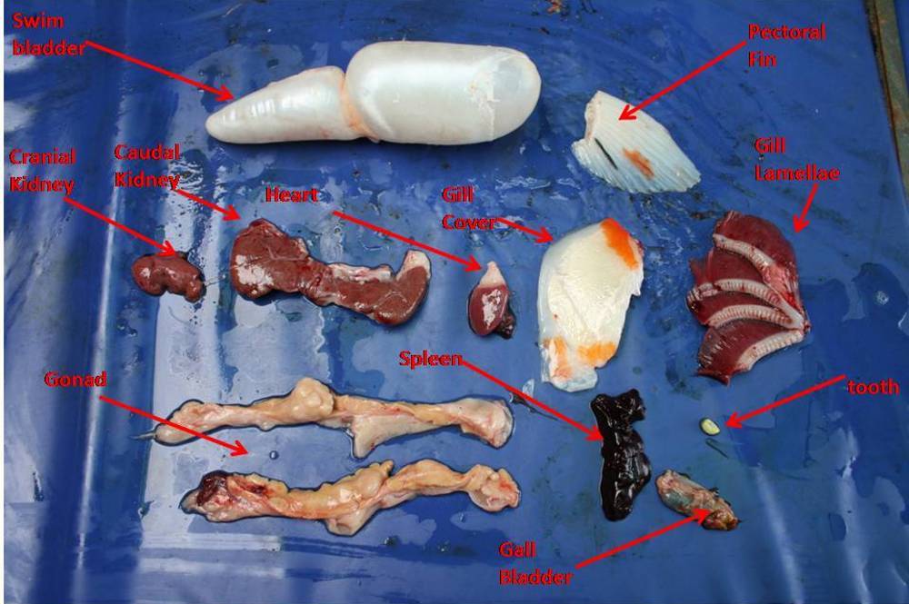
Starting at the front, we have the head or cranial kidney. Like us, the koi has two kidneys but unlike us there is no symmetry in shape or size this is one of the koi’s two kidneys and is just tucked away out of general sight or line of view. the other being the caudal kidney which hangs over the swim bladder like a saddle bag for a horse. This is responsible for osmoregulation. Next we have the swim or gas bladder. This is the organ that controls buoyancy and attitude in water. The swim bladder is a two chambered organ that is joined at the centre by a small duct. On the lower side we have the gut. A koi has no stomach as such just one long tube called the gut which is around 3.5 times the length of the koi’s total; body length. It does however have an oesophagus a large collection chamber for food prior to ist transit down the gut for digestion.
Entangled with and becoming joined/attached to the gut is the liver (arrowed).This is a lighter brown colour than is associated with human animal liver , because the pancreas is integrated into the liver and is not a separate organ is in an animal this the liver is referred to as the hepatopancreas. Also entwined in this mass is the spleen this cannot be mistaken as it is responsible for haemoglobin production thus is a deep red in colour.
At this point I would normally remove the swim bladder first.
Above the swim bladder removed and the caudal kidney. Note: the swim bladder will remain inflated for some time if removed undamaged.
Gonad removed
Ok after this comes the job of sorting the gut from the liver from the spleen and the gall bladder. Don’t worry about the heart it’s safely tucked out of the way for the time being.
This is where it gets messy and gloves are the order of the day. Unfortunately I find it better to gently prise the gut from the liver and spleen and gall bladder
Carefully tease the gut out trying to gently extend the gut tube while doing this using your fingers tease the gut away from the liver tissue. As you progress you will uncover the spleen and the gall bladder these can be removed as a whole and laid to one side, but because the liver and pancreas are inter-twinned
Eventually you will end up with an empty cavity and if you’re lucky the gut laid out as below
The gut laid out which is roughly 3.5X the length of the body
If we switch side now I can show you the empty cavity with the opposite side Gonad and spleen on show this is the dark red organ mid opening arrowed
Next we are going for the heart for this we cut the area that’s left between where the gill cover has been removed and the opening of the main body cavity. Where the stub/base of the pectoral fins has been
Once removed this puts you in the pectoral girdle and access directly to the heart arrowed
The heart can be snipped out and if you have just euthanatized the fish it may still be beating this is an electrical response so don’t be alarmed
Incidentally in the picture you can see the koi’s pharyngeal teeth red arrow
Ok let’s see all the parts and organs laid out

Not included are the liver and gut
Finally I took this a but further this is not normally part of an autopsy of fish but I have included the head section


